x-ray

What is an X-ray?
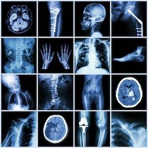
An X-ray is a quick, painless imaging test that allows healthcare providers to view the inside of the body, primarily bones and specific soft tissues. It uses a minimal dose of ionising radiation to view the inside of the body, mainly bones and internal organs, to create images. The denser a structure (such as bones), the whiter it appears on the X-ray film, while softer tissues appear darker.
X-rays are among the most commonly used medical diagnostic tools. They are performed in hospitals, radiology clinics, and emergency rooms.
Who is Suitable for an X-ray?
- Individuals with bone injuries – If a fracture, dislocation, or joint issue is suspected.
- Patients with persistent pain – Especially in the chest, back, joints, or abdomen.
- For people with respiratory issues, X-rays can detect pneumonia, lung infections, and conditions like tuberculosis.
- For those with dental problems, X-rays help diagnose cavities, impacted teeth, or jaw issues.
- People undergoing pre-surgical assessments – To ensure no underlying abnormalities before a procedure.
- Cancer patients – To monitor tumour growth or detect metastasis.
- Individuals with digestive system issues – Some X-ray techniques, such as contrast X-rays, assess conditions in the stomach and intestines.
However, pregnant women should avoid X-rays unless necessary, as radiation exposure can pose risks to the developing baby. In such cases, ultrasound or MRI may be safer alternatives.
Benefits of X-ray
- Quick and painless – The procedure is fast and typically completed within minutes.
- Non-invasive – No need for surgery or incisions.
- Highly effective for bone imaging – Essential for detecting fractures, arthritis, and osteoporosis.
- Aids in early diagnosis – Helps detect conditions before symptoms worsen, allowing early intervention.
- Cost-effective – X-rays are more affordable than imaging methods like MRI or CT scans.
- Low radiation exposure – Modern X-ray machines use minimal radiation while providing clear images.
- Versatile applications – Can be used for bones, chest, abdomen, and dental assessments.
Conditions Diagnosed by an x-ray
X-rays help diagnose a wide range of medical conditions, including:
Bone & Joint Conditions
- Fractures (Broken bones) – Identifies cracks or breaks in bones.
- Dislocations – Determines if a joint has moved out of place.
- Osteoarthritis – Detects joint space narrowing and bone spurs.
- Osteoporosis – Assesses bone density loss.
- Bone infections (Osteomyelitis) – Reveals signs of infection in bones.
Chest & Lung Conditions
- Pneumonia – Detects lung infections and inflammation.
- Tuberculosis (TB) – Identifies lung abnormalities related to TB.
- Lung cancer – Can reveal abnormal masses or tumours in the lungs.
- Collapsed lung (Pneumothorax) – Shows air leakage in the lung cavity.
Heart & Blood Vessel Conditions
- Enlarged heart (Cardiomegaly) – Identifies abnormal heart size.
- Blocked blood vessels – Some X-rays use contrast dye to detect narrowed arteries.
Abdominal & Digestive System Issues
- Swallowed objects – Helps locate ingested foreign objects, especially in children.
- Intestinal blockages – Detects obstructions in the bowel.
- Kidney stones – Identifies calcified stones in the kidneys or urinary tract.
- Gallstones – May be visible on specific X-rays.
Dental & Facial Conditions
- Cavities & Tooth Decay – Helps dentists detect decay between teeth.
- Impacted teeth – Shows misaligned or trapped teeth (e.g., wisdom teeth).
- Jawbone problems – Assists in diagnosing temporomandibular joint (TMJ) disorders.
What Further Information Can an X-ray Show?
X-rays provide detailed images of bones and specific soft tissues, but they can also reveal additional crucial medical information, including:
- Alignment of bones and joints – Helps diagnose joint dislocations, scoliosis, and other musculoskeletal conditions.
- Bone growth or deformities – Useful for detecting abnormalities such as bone tumours, bone infections, or congenital bone defects.
- Foreign objects in the body – Detects swallowed or embedded objects, such as metal, glass, or small plastic pieces.
- Fluid accumulation in the lungs or around organs – Indicates conditions such as pleural effusion (fluid around the lungs) or pericardial effusion (fluid around the heart).
- Soft tissue changes—Certain X-rays, especially those with contrast dye, can highlight abnormalities in the digestive system, kidneys, and blood vessels.
- Arterial blockages – In some cases, X-rays with contrast (angiography) can identify narrowed or blocked blood vessels.
Preparation for an X-ray
X-ray Preparation Summary
- No preparation
- 5-15 mins procedure
- Walk-In service
What to Bring:
- Referral from your doctor – Most X-rays require a medical referral.
- Medicare card or private health insurance details or Worker Compensation details (if applicable).
- Previous X-rays or imaging reports – If a follow-up comparison is needed.
- Comfortable clothing – Depending on the type of X-ray, you may be asked to change into a medical gown.
What to Wear:
- Wear loose, comfortable clothing without metal zippers, buttons, or embellishments.
- Avoid jewellery, piercings, belts, and watches, as metal objects can interfere with imaging.
- Remove bras with underwires if getting a chest or spine X-ray.
How Long Does an X-ray Take?
- Most X-ray procedures take 5–15 minutes.
- More complex X-rays (e.g., contrast X-rays) may take 30 minutes to an hour.
- Additional waiting time may be needed depending on the healthcare facility and workload.
Special Instructions for Certain X-rays:
- Pregnant women – Inform the radiographer before the procedure, as X-rays may need to be postponed or modified.
- Contrast X-rays (e.g., barium swallow, angiography, etc.) – May require fasting for a few hours before the test.
- Spinal or abdominal X-rays – You may need to empty your bladder before the scan.
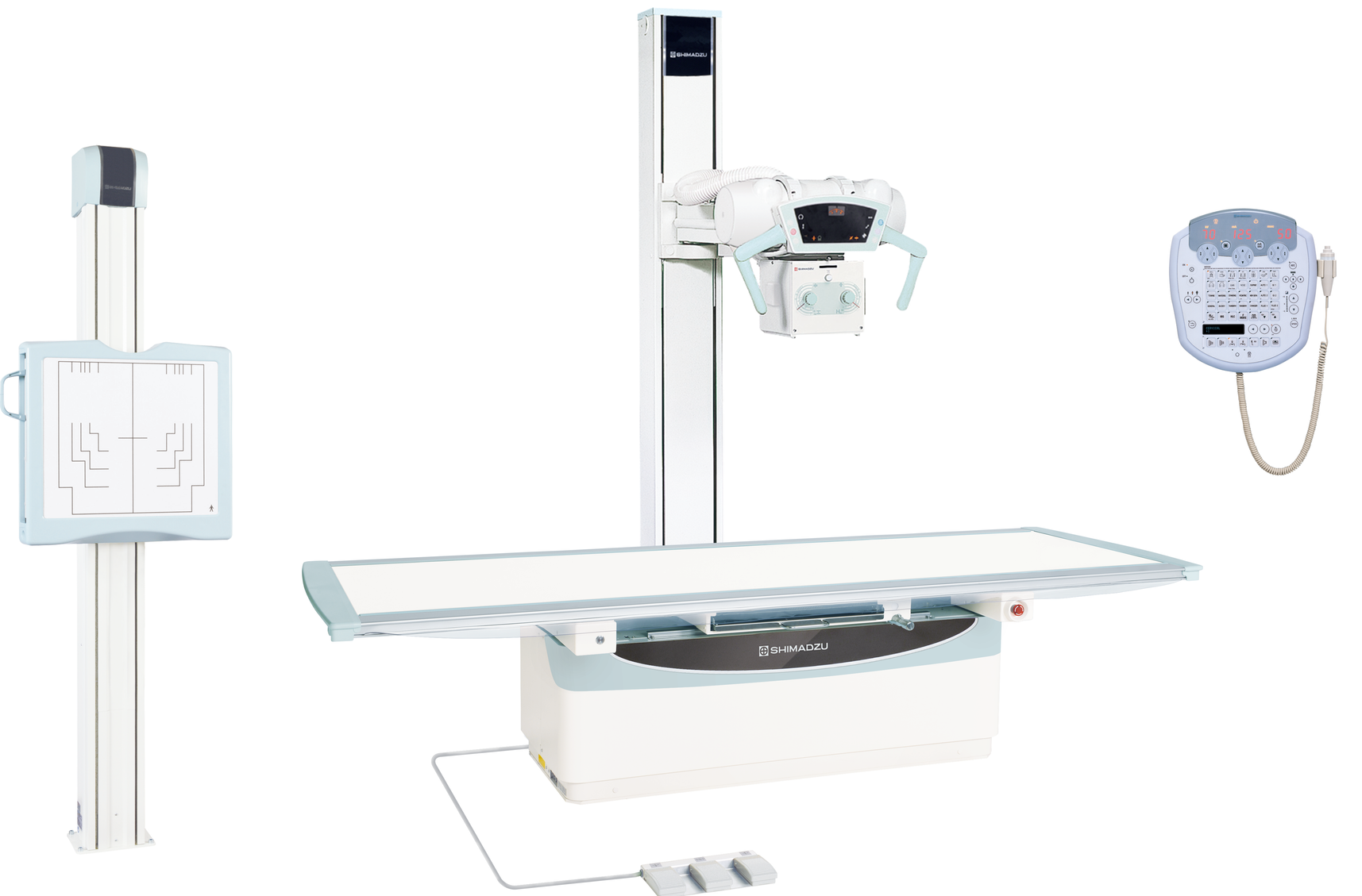
X-ray Procedure
The X-ray procedure is quick and painless, typically involving the following steps:
- Check-in – The radiographer will verify your identity and referral details.
- Clothing adjustment – You may need to change into a medical gown or remove any obstructive clothing/jewellery.
- Positioning – The radiographer will position you depending on the body part being imaged. This may include:
- Standing upright
- Lying down on a table
- Sitting in a specific posture
- Image capture – The radiographer will guide you to stay still while the X-ray machine takes images. You may be asked to:
- Hold your breath for a few seconds (for chest X-rays).
- Change positions for multiple angles.
- Reviewing the images – The radiographer will check if the images are clear. If necessary, additional images may be taken.
The entire process is usually completed within 15 minutes unless additional scans are required.
What to Expect After an X-ray?
Since X-rays are noninvasive and painless, no recovery period is usually needed. However, depending on the type of X-ray, some patients may require specific post-examination instructions.
General Post-X-ray Guidelines:
- You can resume normal activities immediately after a standard X-ray.
- If contrast dye was used (e.g., for a CT or angiogram), you may be advised to drink plenty of water to help flush it from your system.
- If you had a barium swallow or enema, your stool might appear white for a day or two—this is normal.
- Some patients may experience mild discomfort (e.g., soreness from holding a position), but this should resolve quickly.
- Pregnant patients should follow up with their doctor if they are exposed to any radiation.
Getting Your Results:
- Routine X-rays – within 24–48 hours.
- Urgent cases – within 4 hours.
- Your referring doctor will review the images and discuss the findings in a follow-up appointment.
X-ray Prognosis
The prognosis of an X-ray depends on the reason for the scan and the condition being investigated. X-rays provide clear, reliable images that help doctors diagnose various medical conditions, allowing for early intervention and appropriate treatment.
- For fractures and bone injuries – The prognosis is generally excellent if diagnosed early and treated appropriately.
- For lung conditions (e.g., pneumonia, tuberculosis) – Early detection improves treatment outcomes and prevents complications.
- For chronic conditions (e.g., osteoporosis, arthritis) – X-rays help monitor disease progression, allowing for timely management.
- For cancer detection – X-rays (including CT scans) can detect tumours early, improving the chances of successful treatment.
X-ray Risks
A risk commonly associated with X-rays is radiation exposure, which can lead to cancer. However, patients are typically only exposed to a few seconds of low-level radiation in a localised area.
Digital X-rays use less radiation, lowering the probability of any adverse side effects.
On rare occasions, patients may suffer from a severe reaction to the contrast medium and experience:
- Low blood pressure
- Anaphylactic shock
- Cardiac Arrest
What if an X-ray is Delayed?
Potential Consequences of Delaying an X-ray:
- Delayed diagnosis of fractures – A broken bone left undiagnosed may heal incorrectly, leading to long-term complications.
- Progression of infections (e.g., pneumonia, tuberculosis) – Lung infections can worsen and become life-threatening without early detection.
- Missed detection of tumours – If a tumour or abnormal growth is not detected early, treatment options may become more limited.
- Worsening of chronic conditions (e.g., arthritis, osteoporosis) – The disease may progress without a timely diagnosis, causing further pain and mobility issues.
- Unnecessary pain and discomfort – Conditions such as dislocations or kidney stones may worsen, leading to more severe symptoms.
If you have persistent pain, injury, or symptoms, it is best to get an X-ray as soon as possible to avoid complications.
X-ray Costs
Medicare Coverage
- Bulk billed. No out-of-pocket costs.
- A referral from a GP or specialist is required for Medicare rebates.