
ultrasound
What is an Ultrasound?
An ultrasound (also known as sonography) is a medical imaging technique that uses high-frequency sound waves to create real-time images of the inside of the body. It is a non-invasive, painless, and radiation-free procedure commonly used to examine soft tissues, organs, and blood flow.
Ultrasound can be performed on various body parts, such as the abdomen, pelvis, heart, blood vessels, and muscles, providing valuable insights into a person’s health.
Who is Suitable for an Ultrasound?
Ultrasound is suitable for a wide range of patients, including:
- Pregnant Women – Used for monitoring fetal growth, assessing placenta health, and checking for congenital abnormalities.
- Patients with Abdominal Pain – Helps detect gallstones, liver disease, kidney stones, and other abdominal issues.
- People with Suspected Heart Conditions – Echocardiography (is a type of ultrasound) evaluates heart function and structure.
- Patients with Blood Vessel Disorders – Doppler ultrasound checks for blockages, clots, or reduced blood flow in veins and arteries.
- Individuals with Soft Tissue or Muscle Injuries – Identifies sprains, tears, and inflammation in muscles, tendons, and ligaments.
- Women with Gynecological Concerns – Assesses conditions like ovarian cysts, fibroids, and uterine abnormalities.
- Men with Prostate Issues – Helps detect prostate enlargement or abnormalities.
Benefits of Ultrasound
Ultrasound offers several advantages over other imaging techniques, including:
- Non-Invasive & Painless – No incisions or injections are required.
- No Radiation Exposure – Unlike X-rays or CT scans, it is entirely radiation-free.
- Real-Time Imaging – Provides live images, making it helpful in guiding procedures like biopsies.
- Portable & Convenient – Can be performed in clinics, hospitals, and even emergency settings.
- Cost-Effective – More affordable compared to MRI or CT scans.
- Safe for Pregnant Women & Children – No known risks, making it ideal for fetal monitoring.
- Versatile Applications assess multiple organs, blood flow, and soft tissues.
Conditions Diagnosed by an ultrasound
Ultrasound can help diagnose a variety of medical conditions, including:
Abdominal Conditions
- Gallstones
- Liver disease (fatty liver, cirrhosis, tumours)
- Kidney stones
- Pancreatitis
- Appendicitis
Pregnancy & Gynecological Conditions
- Fetal Development & abnormalities
- Ectopic pregnancy
- Ovarian cysts
- Uterine fibroids
- Endometriosis
Cardiac (Heart) Conditions
- Heart valve disorders
- Congenital heart defects
- Fluid around the heart (pericardial effusion)
Vascular (Blood Vessel) Conditions
- Deep vein thrombosis (DVT)
- Aneurysms
- Blocked arteries
Musculoskeletal Conditions
- Tendon injuries (e.g., Achilles tendon tear)
- Muscle tears & sprains
- Joint inflammation (arthritis, bursitis)
Thyroid & Neck Conditions
- Thyroid nodules or goitres
- Lymph node enlargement

What Further Information Can an Ultrasound Show?
Ultrasound provides detailed insights into the body's internal structures, offering more than essential imaging. Depending on the type of ultrasound, it can provide:
- Tissue Structure & Abnormalities – Detects cysts, tumours, inflammation, and infections in organs and soft tissues.
- Blood Flow & Circulation – Doppler ultrasound assesses blood vessels for blockages, clots, or poor circulation.
- Organ Function – Monitors liver, kidney, and heart function by assessing size, shape, and abnormalities.
- Fetal Health & Development – Measures fetal growth, detects birth defects, and evaluates placenta positioning during pregnancy.
- Fluid Build-Up – Identifies fluid accumulation around organs, joints, or in the lungs.
- Soft Tissue & Musculoskeletal Health – Detects tears, inflammation, and structural issues in tendons, muscles, and ligaments.
Preparation for an Ultrasound
Proper preparation helps ensure precise and accurate ultrasound images. Here’s what you need to know:
Ultrasound Preparation Summary
Ultrasound (Thyroid/Neck/Musculoskeletal)
- No preparation
- Walk-in available
- 20 mins procedure
Ultrasound (Abdomen)
- Fasting 8 hours
- 30 mins procedure
Ultrasound (Kidney/Bladder/Pelvis/Obstetric)
- Empty your bladder 2 hours before the scan
- Drink 1 litre of water over 1 hour, and do not empty your bladder
- 30 mins procedure
Interventional procedures (Joints/Spinal injection/biopsy/aspiration)
- Please inform us if you take any blood thinner or have a blood disorder, liver disease or ongoing infection.
- Arrange for someone to pick you up after the procedure
What to Bring
- Referral Letter – From your doctor or specialist.
- Medicare or Private Health Card – if applicable for billing purposes.
- Previous Imaging Reports – If you have had previous ultrasounds, X-rays, or CT scans.
- Comfortable Clothing – Wear loose-fitting clothes for easy access to the scanned area.
What to Wear
- Wear loose, comfortable clothing that allows easy access to the examined area.
- Depending on the type of ultrasound, you may be given a gown to wear.
Diet & Fasting Requirements
- Abdominal Ultrasound – Fast for 6–8 hours (no food, water, or chewing gum) to reduce gas interference.
- Pelvic Ultrasound – This scan requires a full bladder. Empty your bladder 2 hours before your appointment; drink 1 litre of water over 1 hour, and do not empty your bladder.
- Drink 1 litre of water an hour before the test and avoid urinating. A full bladder helps improve image clarity.
- Other Ultrasounds – No special dietary restrictions unless instructed otherwise.
How Long Does an Ultrasound Take?
- Most ultrasound scans take 15–45 minutes, depending on the examined area.
- Some more detailed scans, like echocardiograms or vascular studies, may take longer.
Ultrasound Procedure
Ultrasound is a safe and painless procedure. Here’s what happens step by step:
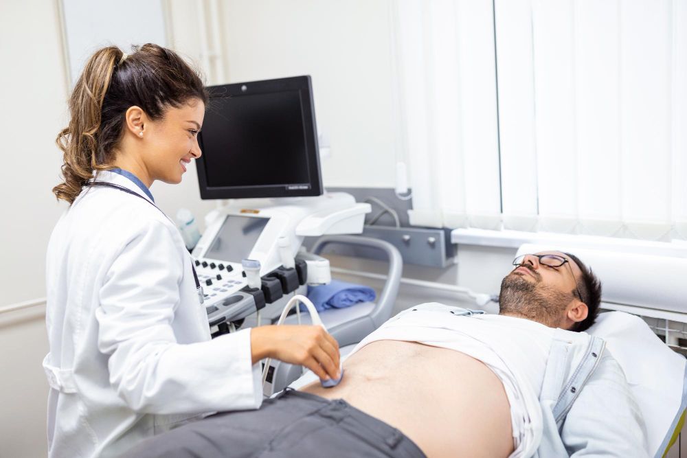
- You will lie down on an examination bed. A water-based gel will be applied to the skin over the examined area, helping the ultrasound probe make better contact with the skin.
- A technician (sonographer) moves the transducer (probe) over the gel-covered area. The device sends sound waves into the body, which bounce back to create images on a screen. You may be asked to hold your breath or change positions for better imaging.
- The technician may listen to blood flow sounds using the ultrasound probe if a Doppler ultrasound is needed.
- After the gel is wiped off, you can resume normal activities. A radiologist reviews the images and sends the results to your doctor.
What to Expect After an Ultrasound?
Immediate Aftercare
- No recovery time – You can return to normal activities immediately after most ultrasounds.
- No side effects – Ultrasound does not use radiation, so no after-effects exist.
- For internal ultrasounds (e.g., transvaginal or transrectal ultrasound), you may experience mild discomfort but can resume normal activities.
Post-Instructions
- If you had a pelvic or abdominal ultrasound with a full bladder, you can use the restroom immediately after the scan.
- For biopsies guided by ultrasound, follow specific care instructions from your doctor.
- If sedation was used (in rare cases), arrange for someone to drive you home.
Receiving Results
- The radiologist analyses the images and sends a report to your referring doctor.
- Results are usually available within 24–48 hours, but urgent cases may receive faster feedback.
- If needed, your doctor will discuss the next steps, including additional tests or treatments.
Ultrasound Prognosis
Ultrasound is a diagnostic imaging tool that helps detect, assess, and monitor medical conditions rather than directly treat them. The prognosis after an ultrasound depends on what the scan reveals:
- Expected Results – No further action is usually needed if the ultrasound shows no abnormalities.
- Abnormal Results – Your doctor will explain the findings and recommend follow-up tests or treatments.
- Monitoring Conditions – Some conditions (e.g., pregnancy, gallstones, liver disease) require regular ultrasounds to track changes over time.
Ultrasound Risks
Ultrasound is considered one of the safest imaging techniques, as it does not use radiation like X-rays or CT scans. However, there are a few potential limitations and risks:
- Mild Discomfort – Some patients may feel slight discomfort, especially during internal ultrasounds (e.g., transvaginal or transrectal ultrasounds).
- Allergic Reaction to Gel (Rare) – Some individuals may have a mild skin reaction to the ultrasound gel.
What if an Ultrasound is Delayed?
Minor Impact (If No Urgent Symptoms)
- A short delay may not significantly affect routine pregnancy scans or minor muscle injuries.
Serious Risks (If Condition is Urgent)
- Undiagnosed Internal Bleeding – A delayed ultrasound may result in life-threatening complications.
- Missed Cancer Diagnosis – Tumors or cysts may grow, reducing treatment options.
- Worsening of Gallstones or Kidney Stones – Delays can lead to more severe pain, infection, or emergency surgery.
- Blocked Arteries or Blood Clots – Delayed Doppler ultrasound may increase the risk of stroke or heart attack.
If your doctor recommends an ultrasound, scheduling it as soon as possible is essential to prevent complications.
Ultrasound Cost
Medicare Coverage
- Bulk billed. No out-of-pocket costs.
- A referral from a GP or specialist is required for Medicare rebates.